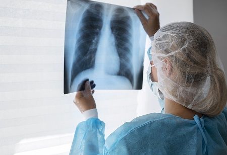
The United States Preventive Services Task Force (USPSTF) recently expanded its lung cancer screening recommendations, which is expected to increase access to screening for women and racial and ethnic minority groups. However, false positives can cause anxiety and unnecessary procedures for patients while increasing costs for the healthcare system. Moreover, efficiently screening many individuals can be challenging depending on healthcare infrastructure and radiologist availability.
At Google, machine learning (ML) models for lung cancer detection have been developed and evaluated for their ability to automatically detect and classify regions showing signs of potential cancer. These models have shown performance comparable to that of specialists in detecting possible cancer. However, effectively communicating findings in realistic environments is necessary to realize their full potential.
In the study titled "Assistive AI in Lung Cancer Screening: A Retrospective Multinational Study in the US and Japan," published in Radiology AI, the effectiveness of ML models in communicating findings to radiologists is investigated. A user-centric interface is introduced to help radiologists leverage these models for lung cancer screening. The system takes CT imaging as input and outputs a cancer suspicion rating using four categories (no suspicion, probably benign, suspicious, highly suspicious) along with corresponding regions of interest. The system's utility in improving clinician performance is evaluated through randomized reader studies in both the US and Japan, using local cancer scoring systems and image viewers that mimic realistic settings. The results show reader specificity increases with model assistance in both reader studies.
To accelerate progress in conducting similar studies with ML models, the code to process CT images and generate images compatible with radiologists' picture archiving and communication system (PACS) has been open-sourced.
Integrating ML models into radiologist workflows involves understanding the nuances and goals of their tasks to meaningfully support them. In the case of lung cancer screening, hospitals follow various country-specific guidelines, such as Lung-RADs V1.1 in the US, to determine follow-up decisions based on CT findings. Radiologists assess patients by loading CT images into their workstations, identifying lung nodules or lesions, and applying set guidelines to make follow-up recommendations.
We use cookies to ensure you get the best experience on our website. Read more...